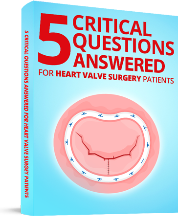
Diagram of Mitral Valve Replacement With Mechanical Valve
I don't know about you...
But when my first cardiologist, Dr. Bad Bedside Manner, told me that I needed open heart surgery due to my defective bicuspid aortic valve... I went into mini-shock.
Then, the stoic doctor, with the red nose, rapidly spewed a flurry of foreign words at me: calcification, leaflets, narrowing valve stenosis, regurgitation, normal ejection fraction, blah, blah, blah, blah.
"Enough medical mumbo-jumbo!" I remember thinking as Dr. Bad Bedside Manner continued to tell me that death was lurking in my future. "Please show me what you are talking about!"
I can't help it. I'm a visual person. I often need to see things to understand them.
For that reason, the cardiologist who I would never see again, grabbed a fake, plastic heart and pointed to the problem area - my aortic valve. He then began promoting open heart valve replacement surgery and the various biological / artificial valve types available to me - pig valves, cow valves, human valves (aka homografts) and mechanical valves.
The discussion referenced above occurred in three minutes. Again, I was in mini-shock.
Considering there are many new patients and caregivers at this website, I figured you may be experiencing what I experienced... A need to see a picture, a diagram or an illustration of a heart valve replacement.
As you can see above, I placed a pretty good diagram which illustrates a mitral valve replacement with a mechanical valve. So you know, I inserted many other images, pictures and illustrations in this website and my book to further help you better understand your heart, your heart valves and the realities of heart valve surgery.
I hope this diagram helps you better understand heart valve replacement surgery.
Keep on tickin!
Adam
Written by Adam Pick
Patient & Website Founder

Written by Adam Pick - Patient & Website Founder
Adam Pick is a heart valve patient and author of The Patient's Guide To Heart Valve Surgery. In 2006, Adam founded HeartValveSurgery.com to educate and empower patients. This award-winning website has helped over 10 million people fight heart valve disease. Adam has been featured by the American Heart Association and Medical News Today.
Comments


Adam's Newest Blogs


New Community Posts
Blog Categories
- Adam's Updates
- Aneurysms
- Aortic Stenosis
- Aortic Valve Repair
- Aortic Valve Replacement
- Atrial Fibrillation
- Before Surgery
- Bicuspid Aortic Valve
- Heart Valve Replacement
- Medical Technology
- Mitral Regurgitation
- Mitral Valve Repair
- Patient Stories
- Pulmonary Valve
- Recovery
- Ross Procedure
- Surgeons & Hospitals
- Tricuspid Valve
Adam's Newest Blogs
Blog Categories
- Adam's Updates
- Aneurysms
- Aortic Stenosis
- Aortic Valve Repair
- Aortic Valve Replacement
- Atrial Fibrillation
- Before Surgery
- Bicuspid Aortic Valve
- Heart Valve Replacement
- Medical Technology
- Mitral Regurgitation
- Mitral Valve Repair
- Patient Stories
- Pulmonary Valve
- Recovery
- Ross Procedure
- Surgeons & Hospitals
- Tricuspid Valve
Heart Hospital Map

Search for heart hospitals that specialize in heart valve treatment.
Surgeon Spotlight
Dr. Christopher Heid is a leading cardiac surgeon at UT Southwestern in Dallas, Texas who specializes in heart valve repair and replacement operations.
New Posts From Our Community

Heather Leigh from
Brooklyn / Nyc
Hi all!! Surgery went well- robotics worked so it was just...
Meet Heather

Adam Pick from
Torrance, California
Hi there! As part of the new community update, all the...
Meet AdamNewest Community Post
Heather Leigh from
Brooklyn / Nyc
Hi all!! Surgery went well- robotics worked so it was just...
Meet Heather
Find Heart Valve Surgeons
Search 1,500 patient-recommended surgeons
Heart Hospital Map
Search for heart hospitals that specialize in heart valve treatment.
Surgeon Spotlight
Dr. Christopher Heid is a leading cardiac surgeon at UT Southwestern in Dallas, Texas who specializes in heart valve repair and replacement operations.