Mitral Leaflet Anatomy, Problems & Pictures
Written By: Adam Pick, Patient Advocate, Author & Website Founder
Page Last Updated: May 16, 2024
I just received an email from Barbara regarding heart valve anatomy, specifically her mitral leaflets.
She writes to me, “Dear Adam – Yesterday, I was unexpectedly diagnosed with severe mitral valve prolapse. Although my cardiologist did his best to explain the problems with my valve, I really could not focus on the words he was saying. I was in shock. There was some discussion about the problems with my mitral leaflet. Can you help me better understand what is wrong with my mitral leaflets? If you have a picture that would be helpful. Best regards, Barbara.”
I can relate to Barbara’s question. When my first cardiologist told me I needed aortic valve replacement, I went numb. My cardiologist talked for another 15 minutes about my diagnosis, but I only understood 10% of the words that exited his mouth. In that moment, words like valve leaflets, dilated heart and left ventricle thickening meant nothing.
Mitral Leaflet Anatomy
Your mitral valve contains two flaps known as mitral leaflets. The leaflets are composed of tissue. The sole purpose of your mitral leaflets is to open and close tightly. This tight seal ensures that blood flows through the heart in one direction, with no blood backflow (known as mitral valve regurgitation) into the heart.

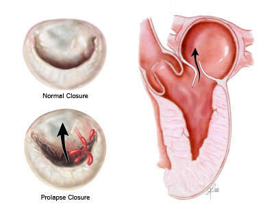
Each heart valve (aortic, tricuspid and pulmonary) has three heart valve leaflets, except for the mitral valve which has two heart valve leaflets. Above, you can see a top-view of the heart with each of the four heart valves visible. In this diagram, the pulmonary and aortic leaflets are open and the tricuspid and mitral leaflets are closed.
Problems With Mitral Valve Leaflets
If mitral leaflets fail to form a tight seal upon closure, the patient could develop heart valve disease. There are several different types of mitral valve disease that result from improper leaflet functioning. Some of these valve disorders are congenital – the patient was born with the problem. In other cases, mitral valve disease developed due to environmental factors (infection, aging) throughout the patient’s lifetime.
Two of the more common issues with mitral leaflets are mitral valve prolapse and mitral valve leaflet calcification. In mitral valve prolapse, the mitral leaflets do not form a tight, smooth seal to prevent the backflow of blood. Instead, the on mitral leaflet sits on top of the other mitral leaflet. You can see a picture below:
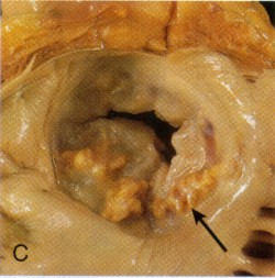
Another common mitral valve disorder results from the build-up of calcium on the heart valve leaflets. Again, calcification of the mitral leaflets could negatively impact the valves ability to open and close. A calcified valve, may narrow and generate a valve disorder known as mitral stenosis. Below you will see a picture of calcified mitral leaflets:
If the patient’s mitral valve leaflet disorders cause short- or long-term issues with heart health, surgery may be required. Surgical approaches for mitral leaflet disorder including mitral leaflet repair and mitral valve replacement.
- Mitral Valve Repair: What Should All Patients Know?
- Mitral Valve Replacement – Patient Facts
- Mitral Leaflets: 7 Important Facts to Know
I hope that helps better explain the anatomy and potential problems with mitral leaflets.
Keep on tickin!
Adam
Written by Adam Pick
- Patient & Website Founder

|
Jennifer From Charlotte says on October 6th, 2008 at 3:48 pm |
|
Fabulous photos!!! Thanks, I will certainly use them in my papers. Jennifer |
 |
|
Felicia says on March 17th, 2010 at 6:55 pm |
|
I recently had mitral valve replacement a month ago. I would like to know if your life is normal and do you have restrictions. I would also like to know how life is living on coumadin for the rest of your life. Can i eat cabbage or greens or spinach ever again. |
|
|
edson luvakubusa says on May 24th, 2017 at 3:59 am |
|
Clinton Luvakubusa |
|
|
L Reynolds says on October 31st, 2018 at 9:08 am |
|
Hello! I have moderate to severe mitral valve regurgitation with both the anterior and posterior leaflets involved. |
|
Adam's Newest Blogs
Adam's Newest Posts


Adam's Video Library
![]()
Learn about heart valve surgery and patient success stories at Adam’s video library.