Surgeon Q&A: “Why Do I Need An Angiogram Before Heart Valve Surgery?” asks John
Written By: Adam Pick, Patient Advocate, Author & Website Founder
Medical Expert: John Grehan, MD
Page Last Updated: June 8, 2025
I recently saw a great question posted at John’s heart valve journal. So you know, John is scheduled for aortic valve surgery on October 17. In his post, John asked, “Since I already know that I have aortic valve stenosis — and need it replaced — why do I have to go through an angiogram?”

I wanted to provide John an expert response to his question. So, I contacted Dr. John Grehan, a heart valve surgeon.
In his email, Dr. Grehan provided several insightful comments that will help John — and maybe all of us — learn more about the use angiograms prior to heart valve surgery.

Dr. John Grehan (Heart Surgeon)
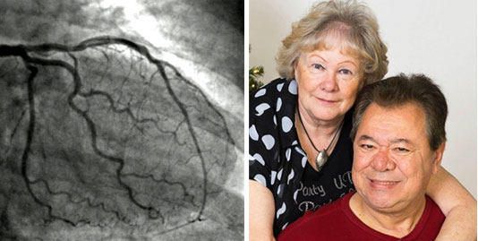
John, thank you for submitting your excellent question regarding the utility of coronary angiography in the pre-operative evaluation of a patient with valvular heart disease. A thorough pre-operative assessment of a patient’s coronary anatomy is an important component of the work up of the patient with heart valve disease. The information obtained from coronary angiography may provide information that will influence the conduct of the planned procedure, alter the surgical approach to the operation, provide a more accurate assessment of the risk the patient is subjected to while undergoing the procedure, and can provide information important in assessing the prognosis of the patient’s condition(s).
The most common use of coronary angiography in the patient with valvular heart disease is to assess the patient for concomitant atherosclerotic coronary artery disease (CAD) that may need to be addressed at the time of heart valve surgery. The presence of significant CAD and valvular heart disease is treated with coronary artery bypass grafting (CABG) at the time of valve repair or replacement surgery. In this setting, coronary angiography demonstrates findings that influence the conduct of the procedure (isolated valve repair/replacement v. CABG/valve repair/replacement), the choice of approach to the procedure (minimally invasive for isolated valve surgery v. standard sternotomy for CABG/valve surgery), the risk profile of the procedure (isolated valve repair/replacement exposes the patient to less risk than CABG/valve surgery), and the long-term prognosis of the patient’s condition (better prognosis in patients with isolated valve disease v. valve disease and CAD). Additionally, in the case of patients with aortic stenosis (such as you), the presence of CAD may even influence the timing of the operation. For example, aortic valve replacement is indicated in patients with significant “surgical” CAD demonstrated on coronary angiography at moderate levels of aortic stenosis. In contrast, in patients with isolated aortic valve stenosis, surgery isn’t indicated until severe trans-aortic valve gradients have been demonstrated.
Another purpose of obtaining pre-operative coronary artery imaging is to define the anatomy of the coronary arteries. A portion of an anatomically “normal” coronary artery “tree” lies in proximity to all of the native heart valves. Therefore, the coronary arteries (or their branches) can be injured, narrowed, or obstructed during valve repair or replacement surgery. Defining the course of the native coronary arteries (and/or any bypass grafts from previous CABG surgery) pre-operatively helps to guide the intra-operative conduct of the planned procedure. Additionally, coronary angiography can demonstrate congenital coronary artery anomalies which can be present in up 5% of the population. The presence of anomalous coronary arteries may influence the pre-operative choice of procedure (and the risk the patient is exposed to) or alter the course of the intra-operative conduct of the operation. Accurate imaging of a patient’s coronary anatomy therefore is important as it provides the surgeon the ability to identify any anatomic variants that may be present specific to each patient prior to bringing that patient to the operating theater. This information may then influence the pre-operative recommendation of the procedure, the choice of approach to the procedure, the intra-operative conduct of the planned procedure, and the early recognition of perioperative complications.
There are two popular modalities for obtaining the necessary images of the coronary arteries. The first, coronary angiography is an invasive procedure and is considered the “gold standard” for assessing patient’s atherosclerotic coronary vascular disease. Coronary anatomy is also demonstrated during this examination allowing for effective pre-surgical planning. The second, more contemporary, imaging modality is CT coronary angiography, a non-invasive radiographic alternative that provides similar information. In my practice I currently utilize both modes of imagining. In patients with multiple risk factors for CAD I recommend proceeding directly to invasive coronary angiography for pre-operative assessment. In younger patients with few risk factors for CAD I recommend proceeding with CT coronary angiography. This defines the coronary anatomy well. If there are no radiographic findings on this examination suggestive of CAD I am comfortable proceeding with valvular heart surgery. If the results of this examination demonstrate concern for CAD however, I recommend follow up examination with invasive angiography to further assess and characterize the patients CAD in order to optimize preoperative planning.
In summary, imaging of the coronary arteries via invasive or radiographic coronary angiography is a necessary component of the pre-operative assessment of the patient with valvular heart disease. The information angiography provides can influence the conduct of the planned procedure, alter the surgical approach to the operation, provide a more accurate assessment of the risk the patient is subjected to while undergoing the procedure, and can provide information important in assessing the prognosis of the patient’s condition(s).
Many thanks to John for posting his question and an extraordinary thanks to Dr. John Grehan for sharing his clinical experience and research with our patient and caregiver community!
Related Links:
- Depression After Cardiac Catheterization?
- Do All Heart Valve Surgery Patients Get an Angiogram?
- Ken’s Cardiac Catheterization: Fear, Pain and Insurance
Keep on tickin!
Adam
|
whatever says on October 8th, 2014 at 2:53 pm |
|
Good question and answer, but makes me wonder why neither angiogram nor angiography were ever given to me as possibilities, but my cardiologists went straight to a heart cath. |
 |
|
Adam says on October 8th, 2014 at 6:41 pm |
|
I just asked Dr. Grehan your question. He responded with the following: “Adam, Angiogram and angiography are synonymous terms. Angiogram is the physical image(s) of the blood vessel(s) being studied. Angiography is the name of the procedure used to image the blood vessels.” “Heart cath is a more generic term. It can (often does) include coronary angiography but will also include direct or indirect blood pressure measurements within the chambers of the heart and a determination of the output of the heart which gives the physician an idea of it’s pumping capabilities.” “Both are appropriate in the pre-operative assessment of patients with heart valve disease.” I hope that helps! |
 |
|
whatever says on October 8th, 2014 at 9:42 pm |
|
Thank you for asking, and it certainly does. |
 |
|
leakyheart says on May 14th, 2016 at 10:37 am |
|
I am wondering is an angiogram considered standard practice, or just a recommended procedure, prior to open heart surgery? My cardiologist is pushing me to get it done, as he was quite surprised my surgeon who I scheduled my open heart surgery with did not recommend it based on his own assessment of my health, but rather offered it just as an option. With the high cost of healthcare these days, I don’t want to risk my health insurance denying this claim because it’s considered a recommended procedure versus a medical necessity, and if it doesn’t show any evidence of coronary blockage or obstruction, i.e., I went through it for nothing. My cardiologist said at my age bracket (I’m 50), health insurance will unlikely challenge covering the procedure whereas if I were younger like in my 20s-30s they would. I’m really torn about whether or not to go through it right now prior to my OHS. |
 |


