
Heart Valve Pictures: Regular & Diseased Heart Valves
Written By: Adam Pick, Patient Advocate, Author & Website Founder
Page Last Updated: June 11, 2025
I'll never forget my second opinion from Dr. Michael Chaikin, a cardiologist in Los Angeles, California.
Dr. Chaikin wanted a real-time echocardiogram done in his office... Unlike many cardiologist, Dr. Chaikin was actually present during the diagnostic test. He studied the monitor as it flashed pictures of my beating heart and pictures of my heart valves. I studied the monitor as well - with great purpose and intent. But, I had no idea what I was looking for.

As it turns out... Eight weeks later, I would find myself having double heart valve replacement surgery via the Ross Procedure. Similar to most patients, I became more interested in heart valve anatomy once I scheduled my open heart surgery with Dr. Vaughn Starnes.
One of the questions I had was, "What does a heart valve really look like?"
I knew my bicuspid aortic valve was a congenital defect. But, I didn't really understand the impact of having two leaflets versus three leaflets. That said, I started looking for heart valve pictures. Here's a quick diagram of a normal aortic valve with 3 leaflets and a bicuspid aortic valve with 2 leaflets fused together.

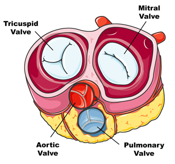
For context, you may want to see a a human heart diagram to understand how the valves are located in the heart. In the diagram below, you can see the position of the aortic valve, the mitral valve, the pulmonary valve and the tricuspid valve. Each valve can experience different congenital and degenerative types of disease. As you can see below, most heart valves have three leaflets, while the mitral valve only has two leaflets.
To start, here is a picture of an actual normal heart valve. You can see how the leaflets are nice and clean with no calcium build-up on the valve. Calcium build-up which can lead to stenosis, or a narrowed valve, this is a very insidious and dangerous disease.

Normal Aortic Valve
Now, let's compare that to an aortic valve with severe disease. As you can see below, the valve cusps are rigid and deformed due to the calcification of the heart valve. This is one of the reasons I needed to have heart valve surgery, my bicuspid aortic valve was very calcified. To learn more about calcified valves, click here.

Calcified Aortic Valve
The more calcified the valve, the more stenotic, debilitating and life-threatening the cardiac condition can be. Here is a chart showing how aortic stenosis can progress over a patient's lifetime.

Aortic Stenosis Progression
As for the mitral valve, the picture below shows a normal mitral valve and several different types of mitral valve disease including degenerative mitral regurgitation caused by mitral valve prolapse, flail leaflets and functional defects. In this diagram, you will note how the blood falls back through the heart due to the improper functioning of the mitral valve. Mitral valve regurgitation is also known as a "leaky heart valve". To learn more about mitral valve prolapse, click here.

It's important to note that mitral valve disease may also be caused due to issues with the mitral valve chordae which are important physical structure that support normal functioning of the mitral valve.

I hope this helped you better understand regular valves and different types of heart valve disease.
Related Links:
Keep on tickin!
Adam
Written by Adam Pick
Patient & Website Founder

Written by Adam Pick - Patient & Website Founder
Adam Pick is a heart valve patient and author of The Patient's Guide To Heart Valve Surgery. In 2006, Adam founded HeartValveSurgery.com to educate and empower patients. This award-winning website has helped over 10 million people fight heart valve disease. Adam has been featured by the American Heart Association and Medical News Today.
Comments


Adam's Newest Blogs


New Community Posts
Blog Categories
- Adam's Updates
- Aneurysms
- Aortic Stenosis
- Aortic Valve Repair
- Aortic Valve Replacement
- Atrial Fibrillation
- Before Surgery
- Bicuspid Aortic Valve
- Heart Valve Replacement
- Medical Technology
- Mitral Regurgitation
- Mitral Valve Repair
- Patient Stories
- Pulmonary Valve
- Recovery
- Ross Procedure
- Surgeons & Hospitals
- Tricuspid Valve
Heart Hospital Map

Search for heart hospitals that specialize in heart valve treatment.

New Posts From Our Community

Cynthia Russell from
Salisbury
So pulmonology says lungs are fine. They think its still the...
Meet Cynthia
Clifford Fava from
Milford, Connecticut
Halfway thru cardiac rehabilitation. This program has sure helped me regain...
Meet Clifford
Patrick Wraight from
Lake Butler, FL
This week is my pulmonologist and pre-op appointment before going in...
Meet PatrickNewest Community Post
Cynthia Russell from
Salisbury
So pulmonology says lungs are fine. They think its still the...
Meet Cynthia
Find Heart Valve Surgeons
Search 1,500 patient-recommended surgeons
Heart Hospital Map
Search for heart hospitals that specialize in heart valve treatment.