Echocardiogram Use For Heart Valve Disease Diagnosis
Written By: Adam Pick, Patient Advocate, Author & Website Founder
Page Last Updated: February 15, 2026
An echocardiogram is a nearly painless procedure that a physician uses to discover exactly how well your heart is transferring blood from one chamber to another.

The test uses sound waves to produce images of your heart. After the test is complete, your doctor will interpret the results and identify any defects in your heart valves or tissues.
How Does an Echocardiogram Help Diagnose Heart Disease?
After listening to your heart with a stethescope, your doctor may determine that you have a heart valve disorder -- often diagnosed as a heart murmur. An echocardiogram is typically ordered both to verify the diagnosis and to reveal the extent of the problem. As the echocardiogram uses sound waves, the technician and physician can view your heart beating.
Typically a standard, two-dimensional echocardiogram takes about 30 minutes to complete. The doctor will then review the report, interpret the film and determine if your heart valves are closing properly. If not, the valves may be narrowed (stenosis) or leaking (regurgitation). The cardiologist will also be able to identify any issues with the structure of your heart (e.g. an enlarged heart or muscle tissue damage).

Different Types of Echocardiograms
Depending on your diagnosis, one of four different types of echocardiogram may be performed.
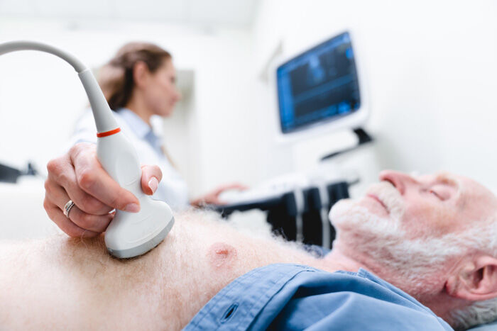
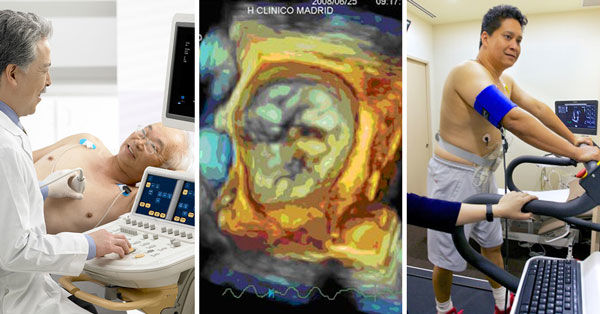
- Transthoracic echocardiogram (TTE) - This is the most commonly used echocardiogram. As shown above, you lie on a table. A monographer, or technician, will use a transducer to project ultrasound beams (high frequency sound waves) to your heart. The transducer is a handheld device which is pressed securely to your chest and moved around so that the technician can record the areas that your doctor has ordered. This test does not require any type of anesthesia or numbing medication since it is painless. However, in the event that your heart cannot be seen through your ribs or lungs, the technician may use intravenous dye to make the images easier to see.
- Transesophageal echocardiogram (TEE) - At times, the cardiologist may decide that he or she would like a view that is unimpeded by ribs or lungs. For that reason, a transesophageal echocardiogram may be requested. In this type of echocardiogram, the transducer is incorporated into an endoscope, which is an instrument used to examine portions of the body from inside rather than outside. You will be given medications to help numb your throat and relax you. The endoscope is passed through your mouth down the esophagus. Once in place, ultrasound is used to produce images of your heart. After the TEE, you may experience some mild soreness in your throat.
- Stress echocardiogram - Some heart conditions cannot be seen while you are resting. These problems only show up when you’re engaged in physical activity. For this reason, your doctor may order a stress echocardiogram. You will either walk on a treadmill or ride a stationary bicycle. An ultrasound will be taken of your heart prior to and immediately after exercising. In the event that you are physically unable to exercise, the doctor may choose to give you medication to make your heart speed up. This makes the heart perform as if you were exercising strenuously.
- Doppler echocardiogram - If your doctor needs to see the direction and speed of your blood flow, he or she may order a Doppler echocardiogram. While a normal TTE or TEE is being performed, the technician also uses Doppler technology to identify certain abnormalities such as valves leaking or poor communication between the atrium and ventricle.
The normal echocardiogram images appear on the screen in black and white, while the Doppler illustrates the blood flow pattern in colored detail.
Recently, I was fortunate to learn about the use of three-dimensional (3D) echocardiograms for diagnosing and treating heart valve disorders at the Heart Valve Summit in Chicago. Here's a video with Christine Wagner, a product application specialist from Philips Ultrasound, that explains the benefits of 3D echocardiograms.
Additional Resources To Help You!
Here's more helpful information about echocardiograms, heart valve disease and our community created to educate and to empower you:


