Echocardiogram Criteria For Mitral Valve Disease
Written By: Adam Pick, Patient Advocate, Author & Website Founder
Page Last Updated: May 10, 2025
Since most people with mitral stenosis and mitral regurgitation are asymptomatic (have no symptoms) until they have had the condition for a while, the first time mitral valve disease is noticed is typically during an annual or routine physical.

Echocardiogram of Mitral Valve Disease
A stethoscope detects sounds of a heart murmur and your physician will order an echocardiogram. Echocardiograms are non-invasive exams that monitor cardiac and valvular function.
Mitral Stenosis
The mitral valve is located between the left atrium and left ventricle. The painless echocardiogram will determine left ventricle (LV) relaxation and also the pressure at which the LV fills. This “resting” phase of your heartbeat is called diastole. The echocardiogram will also allow the sonographer (technician) to measure the area of the mitral valve. The area of the mitral valve, the LV diastolic filling velocity, and the LV filling pressure must be examined to determine the severity of mitral valve stenosis.
LV relaxation is determined by measuring the time necessary for the relaxed LV to obtain the blood from the left atrium (LA) via the opened mitral valve. Your heart is normally very supple and allows the LV to fill quickly during its diastolic phase. When stenosis is present, it becomes more difficult for the diastolic LV to fill with blood. The LA cannot empty as quickly and so maintains pressure longer. If LA pressure remains unchecked, pulmonary edema typically results.
When Doppler is used in conjunction with the echocardiogram, the sonographer can obtain the early velocity at which the LV fills. A healthy heart will have a velocity of 10 cm/sec or higher. Anything less than this indicates impairment of the mitral valve.
Doppler also reveals peak LV filling velocity during diastole. A comparison is then made between them to determine LV filling pressure. LV pressure is expressed in mm/Hg. The chart below reveals the how severe your mitral stenosis is.
The degree of mitral stenosis is determined by the mean gradient of the mitral valve area
- Mild mitral stenosis <5 mmHg >1.5 cm2
- Moderate mitral stenosis 5 - 10 mmHg 1.0 - 1.5 cm2
- Severe mitral stenosis > 10 mmHg < 1.0 cm2
Mitral Regurgitation
The mitral valve damage can either be congenital or because of factors such as calcification. However the damage occurs, as it progresses mitral regurgitation typically results. An echocardiogram must be utilized to determine the severity of the regurgitation.
Although a two-dimensional echocardiogram cannot determine mitral regurgitation, it can identify impairments of blood circulation in the heart and also identify disease development. If mitral regurgitation is to be diagnosed and its severity determined, Doppler must be used.
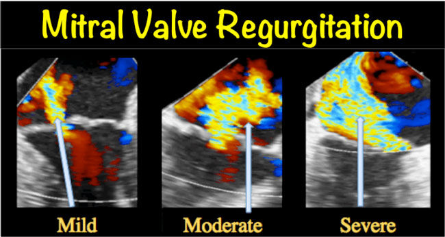
Several different facets of an echocardiogram must be employed to precisely determine the severity of mitral regurgitation. The regurgitant jet size may be determined by color Doppler, the density of the regurgitant jet via continuous-wave Doppler, and finally the inflow of the mitral valve by pulse-wave Doppler. The combinations of these three factors allow the regurgitation volume (RVOL) and the effective regurgitant orifice (ERO) to be determined.
RVOL is obtained as a result of subtracting the mitral stroke volume from the aortic stroke volume. The ERO is obtained by determining the ratio of regurgitant volume to regurgitant jet time-velocity component. Both of these factors, the RVOL and the ERO, can determine the severity of mitral regurgitation.
Mitral regurgitation is mild if RVOL is less than 30ml, and ERO is less than 20mm. Moderate if RVOL is 30-59 and ERO is 20-39. Severe if RVOL is greater than or equal to 60 and ERO is greater than or equal to 40.
In patients who have serious mitral regurgitation but exhibit no symptoms, an echocardiogram is recommended at least annually so that LV size and function may be monitored. Your physician may periodically order a stress echocardiogram to determine how well you can tolerate exercise. This also illuminates how mitral regurgitation and pulmonary pressure react to exercise.
Helpful Resources About Mitral Valve Disease
To further help you learn more about mitral valve disease, here are several new medical posts and patient updates:


